
Latest Findings on Skeletal Structure of the Cell
Scientists at Freie Universität Improve Methods for Imaging the Organization of the Cytoskeleton
№ 245/2015 from Aug 11, 2015
Scientists at Freie Universität Berlin and the Dutch University of Utrecht have developed a method for mapping the structure of the cytoskeleton. The cytoskeleton is the backbone of every cell of the human body. Led by Helge Ewers, a professor of membrane biochemistry at Freie Universität Berlin, and Lukas Kapitein from Utrecht, the researchers succeeded in using light microscopy to resolve so-called microtubules with previously unmatched precision and to examine their arrangement. Microtubules – elongated cytoskeletal polymers assembled from small subunits– are particularly important for the process of cell division and for the transmission of nerve signals in the brain. In the cytoskeleton they form complex structures in bundles in which they, according to the latest measurements by the researchers, approach each other within a few billionths of a meter. The scientists' findings, which were published in the prestigious scientific journal "Nature Communications," may lead to new insight in the development of anticancer drugs.
The arrangement of the cytoskeleton, which consists of microtubules and other dynamic protein polymers such as actin, as well as intermediate filaments, determines the shape of a cell and how its logistics are organized. In particular the thin extensions of nerve cells, that can grow up to several meters in length and transmit signals between different parts of the brain and other body parts, contain tightly bundled microtubules. The mechanism of bundling is important for the growth of new and injured nerve extensions and has therefore been the subject of intense study. Previously this process could merely be investigated using tedious and very time-consuming techniques such as serial section electron microscopy. Due to this technological bottleneck, today's knowledge is based on only a few cells, as systematic functional studies were almost impossible up to now. The new method for optical microscopy described by Ewers' research group rapidly accelerates measurements.
The scientists developed a nanoscopic probe based on antibodies from llamas. Llama antibodies bind to their target with a much smaller domain than those of most other animals, including humans. Probes can be developed from these domains that make it possible to deliver dyes required for microscopy directly to the microtubules. This increases the resolution of super-resolution microscopy by some few, but crucial, nanometers. Eric Betzig, Stefan Hell, and W. E. Moerner received the 2014 Nobel Prize in Chemistry for the development of this process. Ewers and his colleagues now aim to systematically explore the organization of the cytoskeleton by means of these and other newly developed probes.
In 2014 the biochemist Helge Ewers accepted an appointment as full professor of membrane biochemistry at Freie Universität Berlin. In November 2014 he and his group took up their work at the university's Institute of Chemistry and Biochemistry. The scientists in the Ewers Group examine the spatial organization of the proteins of human cells in complex functional units such as synapses. Their research focuses on investigating how millions of different cellular structures can be constructed from the relatively few proteins encoded by our genome. To do so, they combine high-resolution microscopy methods with computer-based image analysis.
Press Image
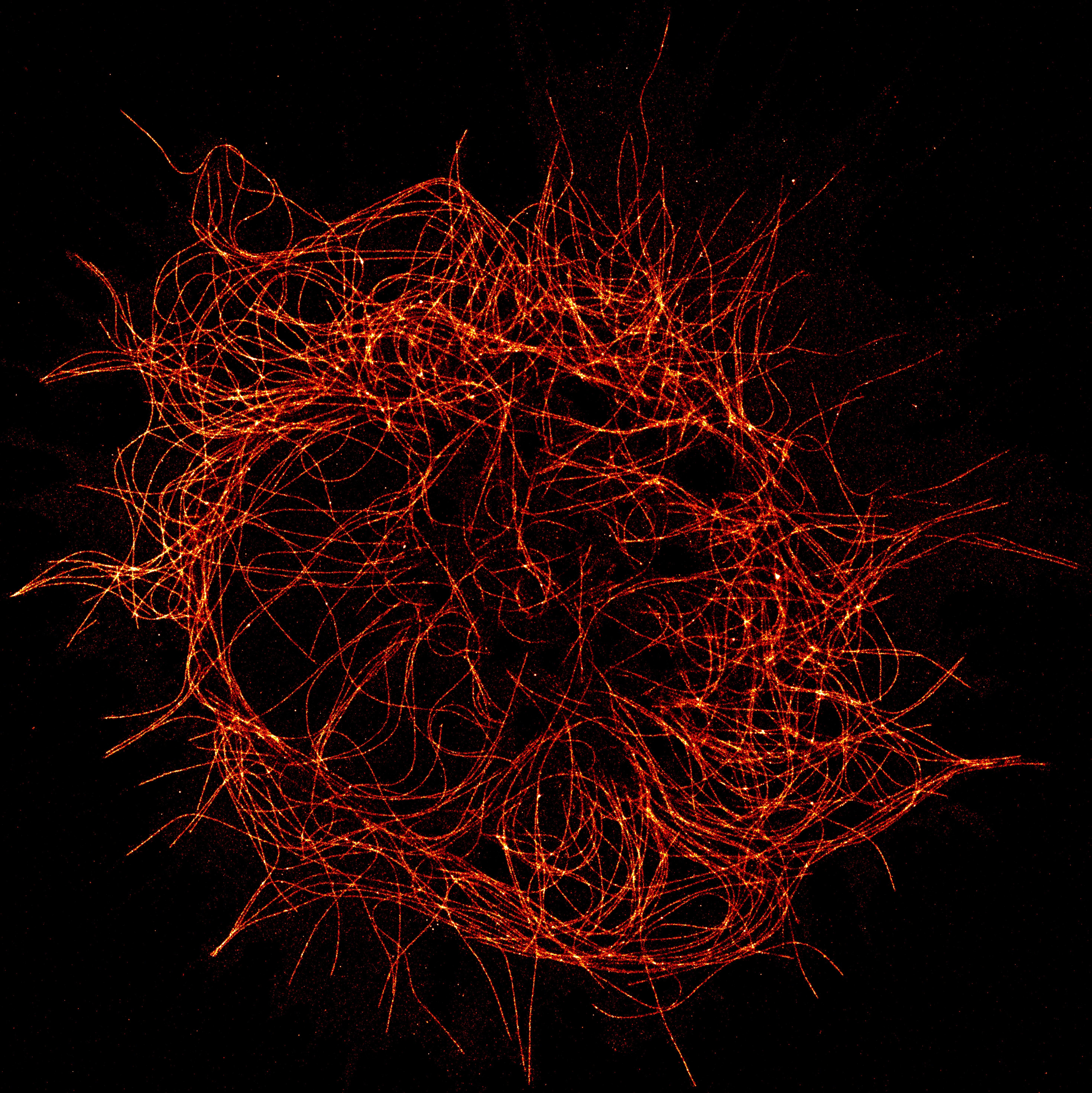
A cytoskeleton, photographed with the method developed by the Ewers Group.
Members of the media may download the image and use it free of charge for reporting in the context of this press release, provided that due credit is given to Ewers Group of Freie Universität.
Further Information
The Publication
Marina Mikhaylova, Bas M. C. Cloin, Kieran Finan, Robert van den Berg, Jalmar Teeuw, Marta M. Kijanka, Mikolaj Sokolowski, Eugene A. Katrukha, Manuel Maidorn, Felipe Opazo, Sandrine Moutel, Marylin Vantard, Frank Perez, Paul M.P. van Bergen en Henegouwen, Casper C. Hoogenraad, Helge Ewers*, Lukas C. Kapitein* (2015): Resolving bundled microtubules using anti-tubulin nanobodies. In: Nature Communications: www.nature.com/ncomms/2015/150811/ncomms8933/full/ncomms8933.html (doi: 10.1038/ncomms8933)
Contact
Prof. Dr. Helge Ewers, Institute of Chemistry and Biochemistry, Freie Universität Berlin, Tel.: +49 30 838-60644, Email: helge.ewers@fu-berlin.de
Link to the Homepage
Information about the Ewers Group: www.bcp.fu-berlin.de/chemie/biochemie/ag/agewers/research