How a Cell's Molecular Knife Is Safely Stored
Scientists in Berlin and Göttingen Discover How a Key Enzyme of the Spliceosome Exerts Its Controlling Function
№ 125/2013 from May 24, 2013
To sustain life, processes in biological cells have to be strictly controlled both in time and in space. Research workers at the Max Planck Institute for Biophysical Chemistry in Göttingen and Freie Universität Berlin have elucidated a previously unknown mechanism that regulates one of the essential processes accompanying gene expression in higher organisms. In humans, errors in this control mechanism can lead to blindness.
DNA (deoxyribonucleic acid) is the carrier of genetic information in all living organisms. Certain regions of DNA, called genes, contain the information that is needed for the assembly of proteins – the molecules that are responsible for most cellular functions. In humans, and in other higher organisms, most genes are built in a mosaic-like fashion – sections that encode the design plans of proteins alternate with so-called non‑coding sections. To produce a protein from the gene that encodes it, first a copy of the gene in the form of RNA (ribonucleic acid, a relation of DNA) has to be produced. From this RNA molecule, the non‑coding sections are removed and the coding sections are spliced together. The result is the so-called “messenger RNA,” the mature RNA that directs protein synthesis. This essential RNA maturation process is termed “RNA splicing.”
The process of splicing is carried out by a highly complex molecular machine termed the spliceosome. Human spliceosomes are built up from protein and RNA molecules. They contain some 170 different proteins and five RNA molecules termed “small nuclear RNAs” (snRNAs). It is currently believed that certain snRNAs represent the tools with which the spliceosome carries out the cutting and joining of RNA sections, turning the messenger RNA's precursor (“pre‑mRNA”) into mature messenger RNA. The proteins of the spliceosome are needed to bring these tools to the right place at the right time, and to set them into operation.
Splicing processes in higher organisms are very highly regulated. In fact, differing patterns of excision and joining of a given pre‑mRNA molecule can lead to any one of a selection of different mature mRNA molecules – all from the same gene. This ability to select the mRNA product according to need is termed “alternative splicing,” and it is thought to be the most important means by which human cells manage to produce a vast spectrum of different proteins from a relatively restricted number of protein-encoding genes.
For every splicing step (removal of a single piece of non-coding RNA), a new spliceosome is assembled on the precursor mRNA molecule. The cutting of the pre‑RNA only takes place once the target splicing site has been identified. The cutting tools of the spliceosome's snRNA are brought into position, but first in an inactive form; they are packed into other components of the spliceosome, rather like a knife that is (initially) kept safely in its sheath. On receiving a particular “start signal,” the molecular knife is drawn and put to use. Until recently, it was only known that a certain protein (known as Brr2) was responsible for activating this molecular knife. Brr2 belongs to a family of enzymes that are called “RNA helicases” due to their ability to separate RNA molecules that are paired with one another (by unwinding the RNA helices, hence the name). In this way Brr2 sets free an snRNA “knife” and allows it to do its job. However, Brr2 also possesses a remarkable molecular architecture, which distinguishes it from other helicases (for details, see “Further publications” no. 1). Until now it was not known how this special architecture is put to use in the cell to regulate the function of Brr2.
Now researchers at Freie Universität Berlin and the Max Planck Institute for Biophysical Chemistry in Göttingen have jointly discovered the mechanism by which this regulation takes place. Sina Mozaffari-Jovin and Cindy Will, from the research group of Reinhard Lührmann in Göttingen, discovered by means of biochemical studies that the helicase activity of Brr2 is inhibited by a particular part of another protein of the spliceosome, Prp8. “Brr2 is, so to speak, held on a short leash by Prp8, preventing it from setting the cutting tools of the spliceosome into action,” explains Reinhard Lührmann.
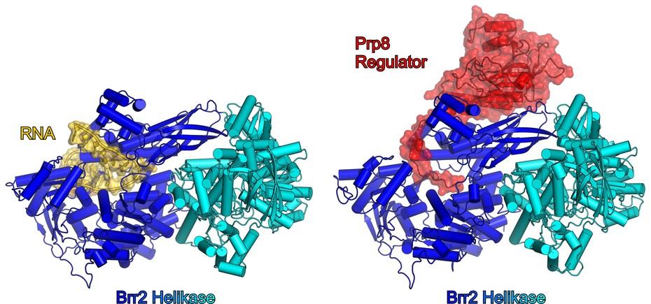
“This prevention requires direct contact between the Prp8 molecule and the helicase Brr2.” Following up on this work, Traudy Wandersleben and Karine Santos, from the research group of Markus Wahl in Berlin, determined the atomic structure of the Brr2 protein in contact with the relevant regulatory portion of Prp8. “To do this we used X‑ray crystallography,” states Markus Wahl. “There are excellent facilities for this kind of research at the BESSY II synchrotrons at the Helmholtz Centre in Berlin, where the necessary specialized instrumentation is available.” The atomic structure explains the biochemical observations in a clear and elegant way: at the end of the Prp8 molecule there is an elongated region of protein that blocks a central channel of the Brr2 helicase and, by doing so, prevents the RNA molecules that are separated by Brr2 from binding to the helicase (Figure 1).
Caption: Figure 1. Left: Model of Brr2 “at work.” Brr2 consists of two similarly structured halves (blue and cyan). An RNA molecule (golden) is shown, lying in a central channel of the left-hand (blue) half of Brr2; this is the molecule to be untwisted. Brr2 can “walk” along this RNA molecule, removing any other RNA molecule paired with it as it goes. Right: Experimentally determined structure of Brr2 with a Prp8 attached to it. The regulatory region of Prp8 penetrates, with its extended structural element, into the channel of Brr2, thus blocking the channel and preventing the RNA substrate from binding. (Photo: Wahl, Freie Universität Berlin)
This region of Prp8, which thus functions like a plug or a stopper, is also significant from a medical point of view. In humans, mutations that lead to changes in this part of Prp8 can lead to a disorder of the eye's retina that can result in blindness. Wahl explains, “The atomic structure of the Brr2–Prp8 complex suggested that these molecular changes in Prp8 might impair its function as a regulator of Brr2. This suggestion was subsequently confirmed experimentally by our colleagues in Göttingen.”
This mechanism is not the only way in which Brr2 is regulated in the spliceosome. The same two research groups recently found an additional regulatory mechanism that controls the Brr2 helicase. Another region of the protein Prp8 binds to the RNA molecules that are to be separated from one another by Brr2, and, by doing so, snatches them away from the enzyme (see “Further publications” no. 2). “The existence of two or more different mechanisms to regulate the same cellular process underlines the importance of the exact timing of this process for the overall process of RNA splicing,” explains the biochemist Lührmann.
Still it remains to be clarified how the various inhibitory mechanisms of the helicase Brr2 work together, and how they are released simultaneously at the right moment – that is, when Brr2 is needed to perform its task of unwinding RNA in the context of the splicing process. The research groups in Göttingen and Berlin have some concrete ideas about how the action of Brr2 might be unleashed, and they plan to pursue these ideas in future research projects. Furthermore, they hope to investigate whether the regulation of Brr2 might also play a part in “alternative splicing.” (mw)
Original Publication
Sina Mozaffari Jovin, Traudy Wandersleben, Karine F. Santos, Cindy L. Will, Reinhard Lührmann, Markus C. Wahl: Inhibition of RNA helicase Brr2 by the C-terminal tail of the spliceosomal protein Prp8. Science, 23 Mai 2013, doi: 10.1126/science.1237515
Other Publications on the Subject
[1] Karine F. Santos, Sina Mozaffari Jovin, Gert Weber, Vladimir Pena, Reinhard Lührmann, Markus C. Wahl: Structural basis for functional cooperation between tandem helicase cassettes in Brr2-mediated remodeling of the spliceosome. Proc. Natl. Acad. Sci. USA 109, 17418-17423 (2012).
[2] Sina Mozaffari Jovin, Karine F. Santos, He-Hsuan Hsiao, Cindy L. Will, Henning Urlaub, Markus C. Wahl, Reinhard Lührmann: The Prp8 RNase H-like domain inhibits Brr2-mediated U4/U6 snRNA unwinding by blocking Brr2 loading onto the U4 snRNA. Genes Dev. 26, 2422-2434 (2012).
Further Information
- www.mpibpc.mpg.de/luehrmann – Website of the Cellular Biochemistry Group, Max Planck Institute for Biophysical Chemistry, Göttingen
- www.bcp.fu-berlin.de/chemie/bc/ag/agwahl – Website of the Structural Biochemistry Group, Freie Universität Berlin
Contact
- Prof. Dr. Reinhard Lührmann, Cellular Biochemistry Group, Max Planck Institute for Biophysical Chemistry, Tel.: +49 551 201-1407, Email: Reinhard.Luehrmann@mpi-bpc.mpg.de
- Prof. Dr. Markus Wahl, Structural Biochemistry Group, Freie Universität Berlin, Tel.: +49 30 838-53456, Email: mwahl@zedat.fu-berlin.de

