
2.3 Million Euros for Nano Research
German Research Foundation (DFG) Approved Funding for New High-performance Microscope at Freie Universität Berlin
№ 288/2016 from Sep 02, 2016
The German Research Foundation has designated 2.3 million euros for a new cryogenic transmission electron microscopy (cryo-TEM) at Freie Universität Berlin. This high-performance microscope will be used by researchers from the Department of Biology, Chemistry, and Pharmacy and cooperating scientists at the Research Center for Electron Microscopy (FZEM) of Freie Universität. The cryo-TEM has evolved in recent years into a powerful tool for high-resolution structural elucidation of natural and synthetic supramolecular aggregates.
Using cryogenic transmission electron microscopy (cryo-TEM), very fine structures in the nanometer range can – while flash-frozen at minus 180 degrees Celsius by liquefied ethane gas – be studied in a fixed aqueous environment. Fixation preserves the natural structures that are formed in solution. Classical electron microscopic preparation methods require drying of the samples, which however, alters the structure and environment of the molecules.
The scientists at the Research Center for Electron Microscopy at Freie Universität Berlin have more than 25 years of experience in working with cryo-TEM and in image processing and 3-D reconstruction techniques using the single particle method. The cryo-TEM to be sponsored by the DFG has a number of new technical aspects, which allow the characterization of low-contrast and radiation-sensitive samples at high resolution. The main use of the new device will be the high-resolution cryo-electron microscopy (including cryo-electronic tomographic) characterization of synthetic supramolecular architectures, synthetic polymers, and organic and inorganic composite nanomaterials. In addition, the scientists will address (structural) biological issues, for example, of proteins, membranes, cell compartments, viruses, or bacteria.
New technical developments that improve cryo-tomographic characterization and increase the typically low contrast enhance the instrumental possibilities for ongoing research collaborative projects, such as the Collaborative Research Centre 765 "Multivalence as chemical organization and action principle" and 958 "Scaffolding of Membranes" as well as planned cooperation research projects dealing with biointerfaces, such as the interaction of biointerfaces with pathogens.
The new cryo-TEM will be operated at the Research Center for Electron Microscopy in cooperation with the Core Facility BioSupraMol. The Core Facility BioSupraMol was funded by the DFG between 2011 and 2015. The purpose of the Core Facility is to advise and support researchers and students at Freie Universität Berlin as well as external researchers in solving complex analytical problems.
The Core Facility BioSupraMol and the Research Center for Electron Microscopy are closely involved in the work of the NanoScale Focus Area at Freie Universität. Since 2008 the NanoScale Focus Area has been a research platform at Freie Universität Berlin that bundles the study of supramolecular interactions as well as bio- and nanomaterials.
Currently, a new building is being constructed at the center of the natural sciences campus at Freie Universität in Berlin-Dahlem, where beginning in 2020, research will be conducted on supramolecular functional architectures at biointerfaces. The new research building will have a special infrastructure and large-scale scientific equipment. Highly specialized laboratories, clean rooms, and low-vibration measuring spaces will be set up.
The purchase of the new cryo-TEM and the construction of the new building are being subsidized jointly by the federal and state governments under the newly amended Article 91 b of the Basic Law for the Federal Republic of Germany. According to this Article, the federal and state governments may cooperate in science and research in cases of national significance. Last year, funds amounting to 37.6 million euros were granted for the new research building.
Press Image
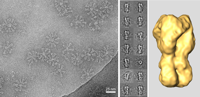
3-D reconstruction of a complex of the viral fusion protein hemagglutinin of the influenza virus with short oligonucleotide chains (aptamers). The structure of this type of complex is intended to help develop optimal inhibitors against viral infection. At left: TEM image of aggregates of the complex ("rosettes"). Center: Typical class sum images resulting from image averaging. At right: Surface representation of the 3-D reconstruction of the complex. Credit: Freie Universität Berlin.
The image may be downloaded by members of the press and used free of charge in connection with reporting on the information in this press release.
Further Information
- Dr. Christoph Böttcher, Scientific Head, Research Center for Electron Microscopy (FZEM), Institute of Chemistry and Biochemistry, Freie Universität Berlin, Tel.: +49 30 838-54934, Email: bottcher@chemie.fu-berlin.de
- Prof. Dr. Rainer Haag, Project Head, Core Facility BioSupraMol, and Deputy Speaker, NanoScale Focus Area, Institute of Chemistry and Biochemistry, Tel.: +49 30 838-52633, Email: haag@zedat.fu-berlin.de